References
What is cfDNA?
Cell-free DNA (cfDNA) consists of short DNA fragments that circulate outside cells in body fluids such as plasma, serum, and urine. These fragments are released during apoptosis and necrosis and can include tissue- or tumor-derived DNA, which makes cfDNA a useful analyte for liquid biopsy, non-invasive prenatal testing (NIPT), and infectious disease diagnostics. [1] Because cfDNA is typically low in abundance and highly fragmented, it is vulnerable to preanalytical variation and genomic DNA (gDNA) carryover from cell lysis. As a result, downstream performance often depends on careful sample handling and extraction choices that preserve short fragments and minimize contamination.
cfDNA vs gDNA: key differences that affect extraction
While cfDNA and gDNA are chemically identical polymers, they represent two distinct biological signatures. gDNA is the stable, high-molecular-weight "archive" extracted from intact cells to establish a patient's inherited genetic baseline. cfDNA, on the other hand, serves as a real-time molecular snapshot, a dynamic marker of physiological or pathological states that is low-abundance and highly fragmented. [2]
Feature | cDNA | gDNA |
|---|---|---|
Biological source | Apoptotic/necrotic cells into body fluids | Inside intact cells |
Physical form & abundance | Short, highly fragmented molecules and low abundance | Long, high–molecular-weight molecules that can be relatively high abundance. |
Pre-extraction stability | Lower. Typically requires timely processing in EDTA tubes (often within hours) to reduce leukocyte lysis and gDNA carryover. | High. Stable for days if refrigerated |
Main use cases | Liquid biopsy, NIPT, minimal residual disease, treatment monitoring, fragmentomics, methylation-based assays | Germline genotyping, inherited disease diagnostics, reference genome analysis, and some tumor tissue profiling |
Risk of cross-contamination | Easily confounded by gDNA released from lysed cells if pre-analytics are not well controlled | Less affected by cfDNA contamination; usually the dominant signal when cells are abundant |
Why is cfDNA Powerful?
-
Often obtainable from plasma, serum, or urine without an invasive tissue biopsy.
-
Allows early detection, molecular profiling, and treatment monitoring for diverse tumor types, as well as applications in NIPT, transplant surveillance, and pathogen detection. [3]
-
Released from apoptotic and necrotic cells, cfDNA captures live signals of tumor burden, therapy response, and minimal residual disease. [4]
Workflow Map
A typical cfDNA workflow includes collection and preservation, separation (plasma/serum or urine supernatant), extraction (spin column or magnetic beads), and QC before PCR/NGS. When results underperform, the root cause is often upstream—especially delays in processing, insufficient stabilization, or inconsistent recovery of short fragments.
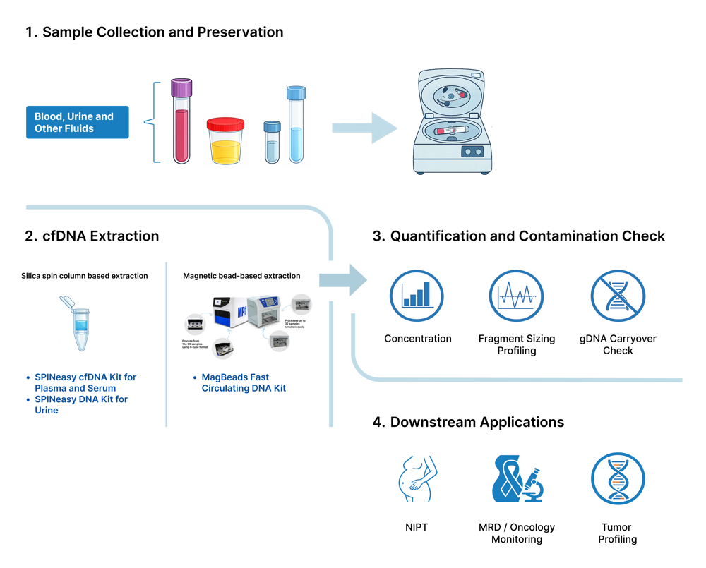
A typical cfDNA workflow includes collection and preservation, separation (plasma/serum or urine supernatant), extraction (spin column or magnetic beads), and QC before PCR/NGS. When results underperform, the root cause is often upstream—especially delays in processing, insufficient stabilization, or inconsistent recovery of short fragments.
Common Issues in cfDNA Extraction and Storage
Poor sample stability
Compared to gDNA, cfDNA is sensitive to preanalytical variation; delays in processing can increase leukocyte lysis and gDNA carryover. Standard EDTA tubes extend whole blood stability somewhat but still require plasma separation within 2–6 hours (ideally <24 hours at 4°C) to minimize leukocyte lysis and gDNA contamination. Specialized stabilization tubes extend this window to days at room temperature but cost significantly more and may contain cell-fixating agents like formaldehyde derivatives that can cross-link DNA, potentially interfering with PCR efficiency or NGS library preparation. [6]
Low Yield
Compared with gDNA, cfDNA’s inherently low and variable yield makes every preanalytical and library‑prep loss critical, directly reducing sensitivity and increasing false‑negative risk in liquid biopsy workflows. Because cfDNA is low and variable in abundance, losses during handling, extraction, or library prep can reduce sensitivity. Some workflows under-recover short fragments if binding and wash conditions are not optimized. [5] Traditional silica-column kits often inefficiently capture crucial short cfDNA fragments (<160 bp), leading to biased libraries that miss biologically important signals.
gDNA Contamination
During improper collection or delayed processing and storage, gDNA often contaminates cfDNA when the cells lyse. White blood cells release gDNA into plasma when stored at room temperature in standard EDTA tubes. This is also observed during shipping where mechanical and thermal stress increases as the preservation tubes are being transported.
Practical solutions for cfDNA stabilization and extraction
Refined Sample Stability and Storage
Degraded or contaminated cfDNA can reduce sensitivity in low-frequency applications such as MRD by increasing background gDNA and diluting low-abundance cfDNA signals. [7] [8]
The Urine Collection and Preservation Kit (Cat. No. 116594000) addresses urine's pre-analytical instability, enabling reliable cfDNA and gDNA workflows even with delayed processing. Both analytes (gDNA/cfDNA) maintain qPCR amplifiability equivalent to fresh samples through ≥30 days RT storage. For plasma-based cfDNA analysis, cfDNA-stabilizing EDTA blood collection tubes like the CelliMAX Cell Free DNA Tube (Cat. No. 091692850) are formulated to minimize leukocyte lysis and preserve nucleic acid integrity during sample collection, transport, and storage. The preservative is formulated to support routine handling without diluting the blood sample. This maintains low baseline cfDNA levels critical for detecting rare ctDNA signals in liquid biopsy, MRD, or NIPT. Together, these products transform sample collection from a time-critical bottleneck into a stable, logistics-friendly platform for sensitive cfDNA workflows.
Optimized cfDNA Recovery
Precision in capturing short fragments is the primary requirement for maximizing cfDNA recovery across diverse sample types. MP Biomedicals addresses this through specialized chemistries that bridge the gap between low-abundance signals and high-sensitivity detection.
For Plasma and Serum
The SPINeasy cfDNA Kit for Plasma and Serum (Cat. No. 116596050) accommodates large sample volumes of up to 10 mL. Its specially formulated Lysis Buffer uses a lysis chemistry designed for efficient processing of plasma/serum inputs and recovery of short cfDNA fragments for PCR/qPCR and NGS workflows. The kit demonstrates superior recovery of short cfDNA fragments compared to competitors, delivering eluates suitable for PCR/qPCR and NGS in liquid biopsy or MRD workflows.

Figure 1: Comparison of extraction efficiency of the SPINeasy cfDNA Kit for Plasma and Serum versus other competitor kits using plasma and urine samples. The SPINeasy kit demonstrates superior recovery of circulating free DNA (cfDNA) as analyzed by Agilent tapestation, highlighting its effectiveness for cfDNA isolation from both plasma and urine matrices.
For Urine Samples
The SPINeasy DNA Kit for Urine (Cat. No. 116593050) isolates both gDNA (pellet, up to 50 mL input) and cfDNA (supernatant, up to 30 mL) using an optimized Binding Matrix. This single-kit solution captures fragmented cfDNA efficiently while supporting paired gDNA/cfDNA analysis, providing a non-invasive silica-column based method for urinary biomarker research.
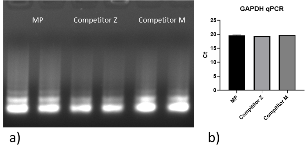
Figure 2: Quality and quantity of cell-free DNA (cfDNA) extracted from a female urine sample using the SPINeasy DNA Kit for Urine, compared with two competitor kits.
a) Agarose gel electrophoresis of cfDNA yield isolated from the female urine sample.
b) Comparison of threshold cycle (Ct) values in qPCR assays using 1 μL of cfDNA isolated from the female urine sample with the SPINeasy DNA Kit for Urine and two competitors. Amplification was performed using SYBR Green technology.
Minimizing gDNA Contamination
To target short fragment loss and enable scalable workflows, the MagBeads Fast Circulating DNA Kit (Cat. No. 117033900) uses magnetic bead technology optimized for high-affinity binding of <200 bp cfDNA fragments from up to 1 mL plasma/serum. Automation-ready for MagFlex-96, MPure-32 aNAP, and compatible with common magnetic extraction workflows, it delivers batch scalability across 96 preps with consistent short-fragment recovery, reduced operator variability through magnetic separation that eliminates centrifugation inconsistencies, minimized gDNA carryover, and concentrated 30–50 µL elutions that maximize input for low-abundance NGS libraries. This bead-based approach complements spin-column flexibility with high-throughput scalability for labs processing dozens to hundreds of cfDNA samples daily.
Explore the MP Biomedicals cfDNA Product Portfolio
Ready to elevate your liquid biopsy?
For low-frequency applications such as liquid biopsy and MRD research, small preanalytical losses can add up quickly. Stabilizing samples early and choosing an extraction approach optimized for short-fragment recovery can improve run-to-run consistency and help protect sensitivity. Use the links below to compare stabilization and extraction options by sample type and throughput needs.
“Cell Free DNA.” ScienceDirect Topics, Elsevier, https://www.sciencedirect.com/topics/medicine-and-dentistry/cell-free-dna. Accessed 8 Jan. 2026. (ScienceDirect)
Qi, Ting, et al. “Cell-Free DNA Fragmentomics: The Novel Promising Biomarker.” International Journal of Molecular Sciences, vol. 24, no. 2, 2023, article 1503. PubMed Central, https://pmc.ncbi.nlm.nih.gov/articles/PMC9866579/. https://doi.org/10.3390/ijms24021503. Accessed 8 Jan. 2026. (PMC)
-
Medina, Jamie E., et al. “Cell-free DNA Approaches for Cancer Early Detection and Interception.” Journal for Immunotherapy of Cancer, vol. 11, no. 9, 2023, e006013. PubMed Central, https://pmc.ncbi.nlm.nih.gov/articles/PMC10496721/. https://doi.org/10.1136/jitc-2022-006013. Accessed 8 Jan. 2026. (PMC)
-
Liu, Shicai, and Jinke Wang. “Current and Future Perspectives of Cell-Free DNA in Liquid Biopsy.” Current Issues in Molecular Biology, vol. 44, no. 6, 2022, pp. 2695–2709. PubMed Central, https://pmc.ncbi.nlm.nih.gov/articles/PMC9222159/. https://doi.org/10.3390/cimb44060184. Accessed 8 Jan. 2026. (PMC)
-
Sathyanarayana, Shivaprasad H., et al. “Standardized Workflow and Analytical Validation of Cell-Free DNA Extraction for Liquid Biopsy Using a Magnetic Bead-Based Cartridge System.” Cells, vol. 14, no. 14, 2025, article 1062. MDPI, https://doi.org/10.3390/cells14141062. Accessed 8 Jan. 2026. (MDPI)
-
Kustanovich, Anatoli, et al. “Life and Death of Circulating Cell-Free DNA.” Cancer Biology & Therapy, vol. 20,no. 8, 2019, pp. 1057–1067. PubMed Central, https://pmc.ncbi.nlm.nih.gov/articles/PMC6606043/. https://doi.org/10.1080/15384047.2019.1598759. Accessed 8 Jan. 2026. (PMC)
-
Peng, Hongwei, et al. “The Impact of Preanalytical Variables on the Analysis of Cell-Free DNA from Blood and Urine Samples.” Frontiers in Cell and Developmental Biology, vol. 12, 2024, article 1385041. https://doi.org/10.3389/fcell.2024.1385041. Accessed 8 Jan. 2026. (Frontiers)
-
Risberg, Bente, et al. “Effects of Collection and Processing Procedures on Plasma Circulating Cell-Free DNA from Cancer Patients.” The Journal of Molecular Diagnostics, vol. 20, no. 6, 2018, pp. 883–892. PubMed Central, https://pmc.ncbi.nlm.nih.gov/articles/PMC6197164/. https://doi.org/10.1016/j.jmoldx.2018.07.005. Accessed 8 Jan. 2026. (PMC)